

Echocardiogram Test for Heart Health: A Complete Guide
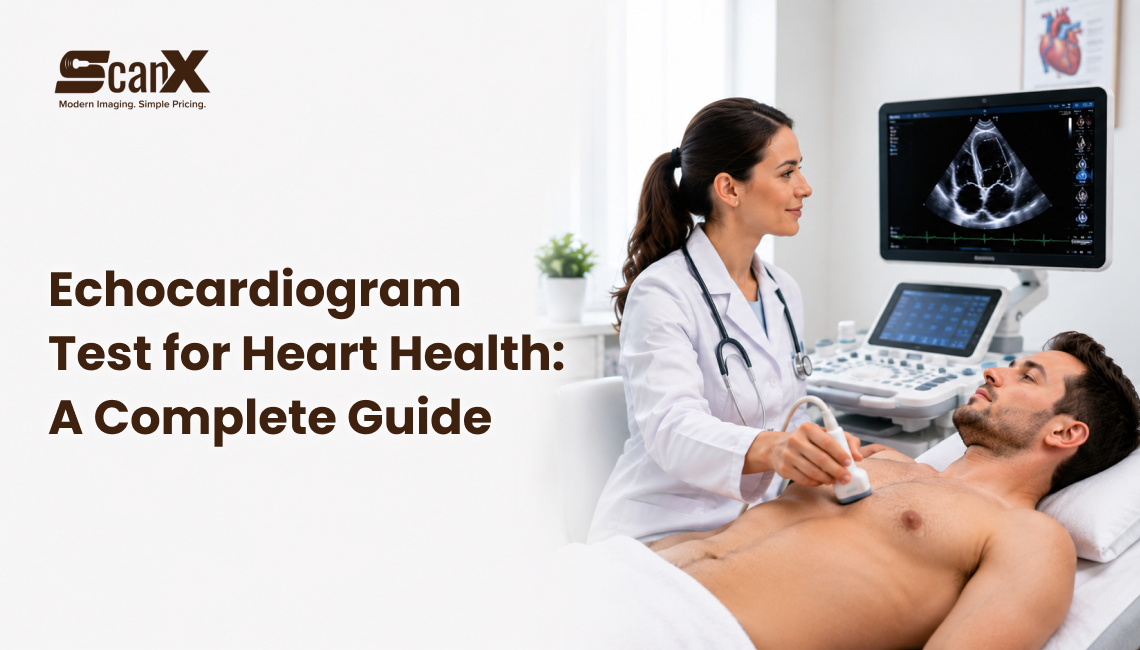
An echocardiogram test is the most essential and widely used heart test that helps doctors see the heart’s structure and how it is functioning. The test uses ultrasound technology. It produces real-time images of the heart.
You can see your heartbeat and your heart pumping blood. This test is safe and non-invasive. It helps diagnose and manage heart conditions. The echocardiogram testis a highly effective and precise assessment of heart health. A heart scan helps doctors assess heart structure, function, and blood flow in real time.
If you have been advised to undergo a cardiac echocardiogram procedure, also known as an echo test, you are not the one. Many people feel really confused or worried when they are advised to undergo an echocardiogram (echo test). The cardiac echocardiogram test for heart is a little scary.
The guide from ScanX is really helpful for understanding your echo test. In this article, we explain what a heart echo test is and why it is very important for detecting heart problems. The echo test for heart gives you a clear picture of your heart’s structure and function. This is very useful for your heart’s structure and function, and the heart echo test helps with that.
If you are getting ready for a heart test or reviewing your test report, this guide will help you understand your heart health test.
You can also read our recent article, “Why Doctors Recommend an Echocardiogram Test,” for a detailed guide and better understanding of the echocardiogram procedure.
What is an Echocardiogram?
An echocardiogram, also known as an “echo,” is a test that uses sound waves to produce real-time images of the heart. It enables medical professionals to ascertain the heart’s chambers, valves, walls, and blood vessels, and to assess blood flow through the heart. This heart chamber evaluation can identify abnormalities and support an accurate diagnosis of heart conditions.
Echocardiograms are painless and noninvasive, with no radiation exposure, making them a safer alternative to other imaging technologies. They are adaptable and can detect a variety of heart diseases, from valve abnormalities to congenital disabilities. This heart scan is safe, painless, and useful for early detection of heart problems. Heart disease screening helps detect early signs of heart problems before symptoms appear.
Get precise echocardiogram test results at ScanX, carefully reviewed by experienced radiologiststo ensure accurate diagnosis and effective treatment planning.
How Does an Echo Test Work for Heart Diagnosis?
The heart imaging test is called an echocardiogram or an echo test. The echo test for heart uses a device called a transducer. An echocardiogram (echo test) is a simple and safe heart imaging test that uses a small device called a transducer. This transducer sends high-frequency sound waves to create images of the heart. These sound waves go back as echoes. The echoes help doctors see how the heart is functioning.
The cardiacechocardiogram is usually performed in a hospital or diagnostic centre. It takes approximately 30 to 60 minutes. So, an echocardiogram procedure is a way to check heart health. It’s a non-invasive procedure. A cardiac echocardiogram is a test used to check heart health.
What are the Types of echocardiogram tests?
Transthoracic Echocardiogram (TTE):
This is the most prevalent heart echo test, where the device is placed on your chest to capture images of the heart.
Transesophageal Echocardiogram (TEE):
In this test, the device is gently passed into the food pipe (esophagus) to obtain clearer, more detailed images of the heart.
Stress Echocardiogram:
This test is executed before and after exercise to assess how your heart functions under physical stress. This heart function assessment can detect early signs of heart problems and guide treatment.
Doppler Echocardiography:
This type of echocardiogram procedure measures the speed and direction of blood flow through the heart.
Why Is an Echocardiogram Done?
Cardiac Echocardiograms (echo tests) play a key role in the diagnosis and monitoring of heart disorders. Doctors typically use an echocardiogram test for heart to:
Check cardiac rhythms or unusual heart sounds
Find heart valve disease, such as narrowing (stenosis) or leakage (regurgitation)
Evaluate the size and operation of heart chambers
Determine inherent (birth-related) heart disorders
Observe cardiovascular health after a heart attack or surgery
Evaluate the ejection fraction to see how well the heart pumps blood
This test helps doctors understand your heart condition definitively and plan the right treatment.
How Does an Echo Test Work in Heart Diagnosis?
An echocardiogram test for heart uses ultrasound technology to capture images of your heart. It helps doctors to check how your heart is functioning. A small device termed a transducer is placed on your chest. This device sends waves into your body. The sound waves bounce off the heart. Come back as echoes. These echoes are used to create moving images of the heart. The echocardiogram test is a painless way to check the heart’s health. The echoes are then used to create images of the heartbeats. A 2D Echo Test provides clear images of your heart to check its structure and function.
A computer then converts these echoes into real-time images, allowing doctors to see how your heart beats and functions. These images can also be saved for future consultation and medical records.
While all echocardiograms use the same basic method, doctors may use advanced techniques to get more detailed information, such as checking blood flow or examining specific parts of the heart.
2D Echocardiogram (2D Cardiac Ultrasound):
This is the most usual type. It creates flat (2D) images of the heart, which are combined to give a complete understanding of its structure.
3D Echocardiogram (3D CardiacUltrasound):
This modern approach provides detailed 3D images of the heart, enabling doctors to examine it from multiple angles with greater accuracy.
Doppler Echocardiography:
This procedure scales the speed and direction of blood flow in the heart and helps evaluate heart function. Heart function assessment helps doctors check how well your heart pumps blood.
Strain Imaging:
This method presents how well your heart muscles contract and relax, helping detect early signs of heart problems.
Contrast Echocardiogram:
In this test, a particular contrast dye is inserted into a vein to highlight heart structures and improve image clarity.
An echocardiogram (echo test) does not use radiation, making it a safe and preferred choice for heart imaging. Unlike X-rays and CT scans, which use minor amounts of radiation, a heart echo test uses harmless sound waves to create images of your heart. The 2D Echo Test is a risk free and non-invasive way to detect heart problems early.
How to Prepare for a Heart Ultrasound Test
Preparation for an echocardiogram (echo test) depends on the type of test:
Transthoracic Echocardiogram (TTE):
No particular preparation is needed. You can eat, drink, and in take your regular medications.
Transesophageal Echocardiogram (TEE):
Avoid drinking anything for 6 to 8 hours before taking the test. If the doctors are going to give sedation, you need to find someone who can drive you home after the test.
Stress Echocardiogram:
Wear comfortable clothes and exercise-appropriate shoes. Avoid caffeine and heavy foods before the test to ensure accurate results.
Proper preparation helps ensure accurate and reliable echocardiogramprocedure results.
During the echocardiogram (echo test), cautiously obey the technician’s instructions to help capture clear and precise images of your heart
How Doctors Interpret Echocardiogram Test
Cardiologists review echocardiogram (echo) findings to check whether your heart is functioning smoothly. These cardiologists recommended tests to provide accurate insights for better heart care and treatment planning.
Normal echocardiogram values include:
Ejection Fraction (EF): A range of 55–70% shows that your heart is pumping blood properly
Heart Chamber Size: Should be normal based on your age and body size
Valve Function: Heart valves should open and close properly without leakage or narrowing
These results help doctors understand your heart condition and determine if any medication is required.
What Do Abnormal Echocardiogram Results Mean?
Abnormal echocardiogram results may indicate heart disorders such as heart failure, valve issues, blood clots, or fluid around the heart. Your doctor will interpret these results and may recommend further tests or appropriate treatment if needed.
What Are Normal EchocardiogramResults?
Normal echocardiogram results indicate that your heart is functioning well. They confirm that your heart is pumping blood adequately and that there are no blood clots or abnormal structures in the heart.
ScanX Expert Note: Echocardiogram Insights
An echocardiogram (echo test for heart) is a common and safe heart test that provides information about your heart’s structure and function. If your doctor suggests an echocardiogram, you can ask about the type of test and what to anticipate during the procedure.
It’s normal to feel a little nervous before diagnostic testing. At ScanX, our trained experts ensure you feel comfortable and relaxed throughout the echocardiogram process. It's a quick and painless heart health test, and it provides important insights to help monitor and protect your Cardiovascular health.
At ScanX, we provide affordable, transparent, and patient-centred ultrasound imaging servicesin Dallas and Fairview, ensuring high-quality care at competitive prices.
Our Patient Referrals program makes it simple for patients to access fast, reliable, and cost-effective imaging services without hassle.
Conveniently located in Dallas, ScanX proudly serves patients from Arlington and nearby cities, including Rockwall, Rowlett, Irving, Flower Mound, McKinney, Frisco,Garland,Plano, Allen, and surrounding areas.
You can also explore our article, “7 Reasons Doctors Recommend a Carotid Doppler Ultrasound Test,” for a quick and clear comparison between a carotid ultrasound and a CT scan.
Conclusion
An echocardiogram test is a crucial diagnostic procedure for evaluating heart health. By understanding what an echocardiogram includes, how to prepare, and what the results mean, patients can access the procedure with confidence. Always discuss the findings with your doctor to interpret them and determine the best course of action. This heart disease screening supports timely diagnosis and better cardiovascular health management.
Explore our comprehensive range of affordable imaging services at ScanX, including thyroid, liver,kidney, gallbladder, and abdominal ultrasounds. We also offer specialised scans such as hernia imaging, FibroScan liver elastography, carotid ultrasound, and AAA screening.
Bookyour Echocardiogram test for the heart today and take a proactive step toward better heart health and overall well-being.