

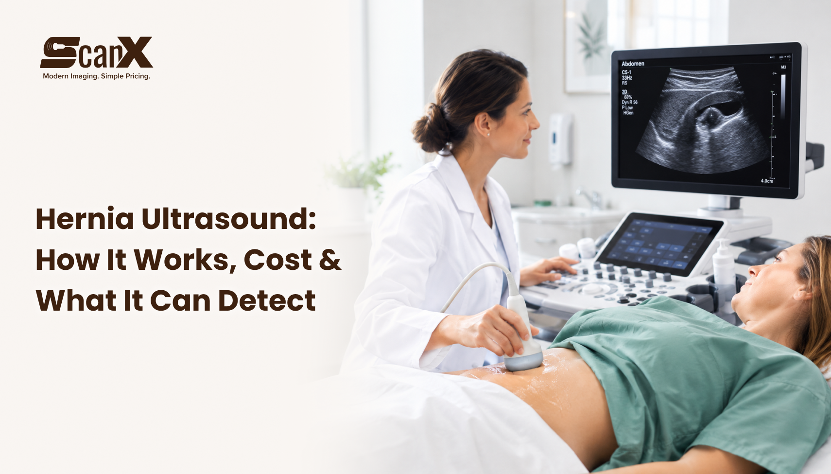
Hernia Ultrasound is a safe and painless test to detect hernias
A hernia ultrasound is a quick, safe, and painless scan that uses sound waves to produce pictures of the tissues and muscles inside your body. It is commonly used to check the abdomen and groin, where hernias are most common.
A hernia emerges when an organ or tissue forces through a weak spot in the muscle wall. This can cause a bulge, pain, or discomfort, especially when you cough, lift, or strain.
A hernia ultrasound helps doctors confirm whether you have a hernia, see the size and exact location of the hernia, and plan the appropriate treatment, including surgery if needed.
The test is non-invasive, uses no radiation, and takes only a few minutes. It is safe for people of all ages, including children and pregnant women.
Want to know more about ultrasound scans? Read our latest post, “Thyroid Ultrasound Test: Why It’s Done, Cost & Normal Results Guide.”
How Does a Hernia Ultrasound Work?
Can an ultrasound detect a hernia? Yes, it clearly shows hernias in the groin and abdominal wall, including small ones that are hard to feel during a physical exam. During a hernia ultrasound, a technician gently moves a small handheld device termed a transducer over the skin of the area where a hernia is suspected, usually the abdomen or groin. The transducer sends sound waves into your body, and these waves bounce back to form clear images of your muscles and tissues.
These images show whether an organ or tissue, such as part of the intestine, has pushed across a weak spot in the muscular layer. This is what causes a hernia. An abdominal wall hernia ultrasound is a quick, painless, radiation-free scan that shows the size, location, and contents of a hernia.
A groin ultrasound for hernia checks the inguinal and femoral areas using sound waves, showing whether tissue or intestine is pushing through the muscle wall, even if the bulge is small or comes and goes.
Inguinal Hernia Ultrasound
To check for an inguinal hernia, the sonographer places the transducer over your groin. You may be asked to stand, cough, or strain so the pressure in your abdomen rises and the hernia becomes easier to see. The ultrasound can show part of the intestine pushing through the inguinal canal and help tell a direct inguinal hernia from an indirect one. This difference is important because it helps the surgeon plan the right repair, if surgery is needed.
Can an Ultrasound Detect an Inguinal Hernia?
Yes. An ultrasound can detect an inguinal hernia, and it is a safe, painless, radiation-free way to check the groin. During the scan, the sonographer uses live imaging while you lie down, stand, cough, or bear down. This raises the pressure in your abdomen and helps the hernia show up on screen. The scan can also tell a direct inguinal hernia from an indirect one and help rule out other groin lumps, such as swollen lymph nodes, lipomas, or hydroceles.
Curious how IVF monitoring works? Check out our latest article, “Understanding IVF Monitoring Ultrasounds: A Patient’s Guide.”
Why Choose a Hernia Ultrasound?
Hernia ultrasound accuracy is high for most groin and abdominal wall hernias, especially when the scan is done while you stand, cough, or strain, though small or hidden hernias may sometimes need an MRI or CT scan to confirm.
Non-invasive: No surgery, needles, or cuts are needed.
Quick: The scan usually takes only 15 to 30 minutes.
Radiation-free: It is a safe imaging test for everyone, including children and pregnant women.
Painless: You may feel only light pressure from the transducer.
What Are the Benefits of a Hernia Ultrasound?
Using an ultrasound to detect a hernia is a quick, painless, and radiation-free way to see its size, location, and contents, without surgery or needles. A hernia ultrasound offers various significant benefits for diagnosing and treating hernias early and safely. Here is how it can help:
Early Detection of Hernias
A hernia ultrasound can clearly confirm whether you have a hernia. Small hernias or hernias with mild symptoms are often hard to find during a physical exam, but an ultrasound shows them clearly. It reveals if a hernia is present, where it is located, and how big it is. Finding a hernia early means you can get treatment sooner, which helps stop it from getting worse.
Better Treatment Planning for Hernias
Once a hernia ultrasound confirms a hernia, doctors can decide the best treatment plan for you. Some hernias need surgery to repair the weak muscle wall. Others are small and cause no symptoms, so they can be watched over time. The ultrasound for hernia provides doctors with a clear picture of the hernia, which helps them decide whether you need surgery right away or can safely wait and monitor it.
Tracking Hernia Changes Over Time
If you have a small hernia that does not need surgery immediately, a hernia ultrasound can be used to keep an eye on it. Regular scans let doctors see if the hernia is getting bigger or causing new symptoms. This helps them decide if you may need surgery in the future.
Safe, Painless, and Non-Invasive Imaging
A hernia ultrasound is absolutely non-invasive. There is no surgery, no radiation, and no recovery time. The scan is painless and safe for everyone, including people who need repeat scans to keep track of a hernia over time.
Fast and Easy Hernia Scan
A hernia ultrasound is quick and simple. The scan commonly takes less than 30 minutes, and results are ready quickly. This helps doctors decide on treatment without delay, which matters most if you have pain or discomfort from a hernia.
When to Get a Hernia Ultrasound
Your doctor may recommend a hernia ultrasound if you have:
An evident bulge in the abdomen or groin region.
Pain or distress when bending over or lifting something heavy
Swelling or tenderness in the abdomen or groin
Digestive problems, such as trouble passing stools
A hernia ultrasound is a useful tool for finding and managing hernias. ScanX recommends it because it is a safe, painless, and non-invasive way to confirm a hernia, guide treatment decisions, and track changes over time. By finding a hernia early and helping doctors choose the right treatment, a hernia ultrasound can help avoid complications and lead to better health outcomes.
When a Hernia Becomes an Emergency
If you notice any of these symptoms, it is important to see a surgeon who specializes in hernia repair. Finding a hernia early and getting treatment quickly can help prevent complications and improve your living standards. For more details, read our guide on hernia symptoms and diagnosis: when to seek surgery in Dallas.
How Much Does Hernia Ultrasound Cost?
The national average cost of hernia repair surgery ranges from approximately $3,057 to $14,304. The final price depends on the type of surgery, the type of anaesthesia used, the surgeon’s experience, and your location. The hernia ultrasound cost breakdown by surgery type shows what a typical person might pay without insurance, including copays, deductibles, and other out-of-pocket costs. The cost of hernia surgery depends on the type of procedure you choose. Open hernia surgery costs about $4,500 on average, with prices ranging from roughly $3,100 to $7,600. Laparoscopic hernia surgery averages around $4,900, with a typical range of about $3,100 to $8,400. Robotic hernia surgery is the most expensive option, averaging close to $8,400, with prices ranging from about $5,700 to $14,300.
Will Insurance Pay for Hernia Repair?
Most health insurance plans cover hernia repair surgery as long as it is medically necessary. Insurance usually pays for the surgeon’s fee, anaesthesia, and facility charges. Even when surgery is covered, you may still need to pay deductibles, copays, or coinsurance.
Not sure whether to use insurance or pay directly? Read our guide to self-pay vs insurance pricing for ultrasounds.
How a Hernia Ultrasound Helps Prevent Complications
A hernia ultrasound can help doctors spot significant health problems, such as damaged or dead tissue, before they get worse. During the scan, a small probe sends sound waves into the body to create clear images of the tissues and the area around the hernia. If you have pain, discomfort, or other hernia symptoms, your doctor may suggest this scan to see exactly what is going on. Finding a hernia early means you can get the right treatment sooner, which lowers the risk of complications from the hernia itself or from repair surgery.
Why Choose ScanX for Your Ultrasound?
When you have a sudden bulge, pain, or swelling, you need clear answers fast, without long hospital waits or high bills. ScanX makes it simple. We offer affordable self-pay ultrasounds with prices you can see before you book. Every scan is done by a certified sonographer and read by a board-certified radiologist, so you get accurate results and peace of mind. Your secure results are ready within 24 to 48 hours, so you and your doctor can plan the next step, including a surgeon visit if needed.
Not sure what that bulge means? Don’t wait for your symptoms to get worse. Book your ultrasound for hernia at ScanX today and get the answers you need. Book your appointment here.
Conclusion
Hernia Ultrasound at ScanX with clear answers for every type of Hernia. A hernia ultrasound is a key tool for finding and managing hernias. It gives real-time, non-invasive images that help doctors confirm a hernia, check how serious it is, and choose the right treatment. Whether you have an inguinal, femoral, umbilical, or incisional hernia, an ultrasound gives you the clarity to make confident healthcare decisions.
Finding a hernia early can improve your outcome. Following your doctor’s advice and getting regular follow-up scans can help manage the hernia and prevent complications. If you or a loved one has a bulge, pain, or swelling, don’t wait. Book a hernia ultrasound at ScanX in Dallas & DFW for $124, with radiologist-reviewed results in 24 to 48 hours. Book your appointment here.
Searching for a hernia ultrasound near me? Choose a certified imaging centre with clear self-pay pricing, like ScanX, so you know your cost before you book.
ScanX in Dallas welcomes patients from nearby cities like Arlington, Rockwall, Rowlett, Irving, Flower Mound, McKinney, Frisco, Garland, Plano, and Allen. Book a hernia ultrasound close to home.
Searching for a trusted ultrasound provider in Dallas or Fairview? ScanX delivers clear, upfront pricing and patient-first care. Use our Patient Referrals page to get your ultrasound imaging services without the hassle. We offer liver, kidney, gallbladder, and abdominal ultrasounds, plus hernia scans, FibroScan liver elastography, carotid ultrasounds, and AAA screening. Schedule your hernia ultrasound today.
FAQs
1. What Happens During a Hernia Ultrasound?
A hernia ultrasound is a safe, easy, pain-free, and non-surgical scan that uses sound waves to look at your muscles, abdominal wall, and any bulging tissue or bowel. It is usually done on the groin, lower abdomen, or belly button area. During the scan, you will first lie down on the exam table, and you may be asked to stand later, since gravity can help show smaller or hidden hernias. A warm liquid gel is applied to the skin, and the sonographer moves a small handheld device called a transducer over the area. Light pressure may be used to check if the bulge can be pushed back in. You may also be asked to cough, bear down, or lift your head so the hernia becomes easier to see. If needed, color Doppler imaging checks blood flow to make sure the tissue is healthy.
2. Do I Need an Ultrasound for a Suspected Hernia?
Not always. If a doctor can see or feel a clear bulge, especially when you stand, cough, or strain, a physical exam is usually enough to diagnose a hernia. Guidelines such as Choosing Wisely advise against routine ultrasound for obvious inguinal or umbilical hernias, since it can add cost and delay without changing treatment. Your doctor may suggest a hernia ultrasound if the exam is unclear, if you have symptoms but no visible bulge, or if the hernia is small or hidden. It is a safe, painless, and radiation-free scan that can be done standing or while straining to bring out the hernia. A CT scan is usually reserved for complex or emergency cases.
3. What Is the Difference Between a Hernia Ultrasound and a CT Scan?
A hernia ultrasound uses sound waves to create live images and involves no radiation. A CT scan utilizes X-rays and a computer to produce comprehensive cross-sectional pictures of the abdomen. Both can find a hernia, but they work differently and are used in different situations. Technology and safety: An ultrasound can be done while you stand, cough, or strain so that doctors can watch the tissue move in real time. It is safe for all ages, including pregnant women, and can be repeated when needed. A CT scan is done while you lie flat, uses ionizing radiation, and sometimes needs contrast dye through an IV. When each is used: A hernia ultrasound is often the first test for simple or groin hernias, or when you have pain but no clear lump. It can also catch small or on-and-off hernias that may slip back in when lying down. A CT scan is usually reserved for large, complex, recurrent, or incisional hernias, and for emergencies such as severe pain, bowel blockage, or strangulation.
4. Can an ultrasound detect a small hernia?
Yes. A hernia ultrasound can find small or hidden (occult) hernias, especially in the groin and abdominal wall. During the scan, you may be asked to stand, cough, or bear down so that a small bulge pushes out and becomes easier to see. The ultrasound shows the gap in the muscle wall and any fat or bowel pushing through. It can also help tell a hernia apart from other lumps, such as a swollen lymph node, a lipoma, or a fluid collection. Finding a very small or on-and-off hernia depends on the sonographer’s skill and on whether the hernia shows up during the scan. Extra body fat can make small defects harder to see. Deep internal hernias, such as a hiatal hernia, are better seen with a CT scan or MRI. If your ultrasound is negative but you still have pain or pressure, your doctor may suggest an MRI for a closer look.
5. Is a Hernia Ultrasound Painful?
No. A hernia ultrasound is not painful. It is a non-invasive scan that uses sound waves, with no needles and no radiation. The sonographer spreads a cool, water-based gel on your skin and glides a small handheld probe over your groin or abdomen. You may be asked to stand, cough, or bear down so the hernia becomes easier to see.